This Rice University Professor Developed Cancer-Detection Technology

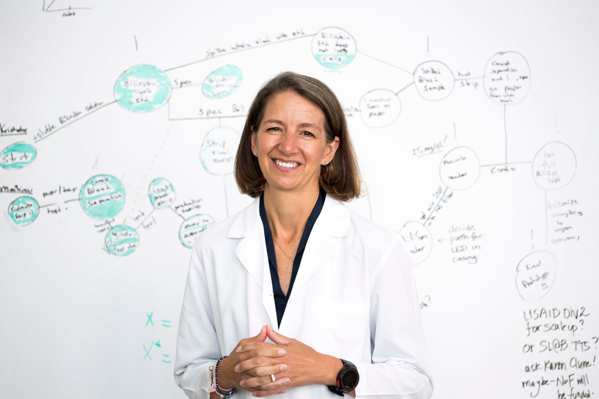
Rebecca Richards-Kortum has spent most of her 30-year career developing technology to help improve medical care in underserved communities worldwide. Among her achievements: She invented an inexpensive, battery-operated optical imaging system that can detect premalignant tissues-no biopsy required-to help prevent oral and cervical cancer.
Richards-Kortum is a professor of bioengineering at Rice University, in Houston, and codirector of the Rice360 Institute for Global Health Technologies, which is developing affordable medical equipment for underresourced hospitals. Her team created a suite of low-cost medical devices, the NEST360 newborn tool kit, to improve neonatal health in sub-Saharan Africa.
Rebecca Richards-KortumEmployer
Rice University in Houston
Title
Director of the Rice360 Institute for Global Health Technologies
Member grade
Senior member
Alma maters
University of Nebraska-Lincoln; MIT
For her contributions to optical solutions for cancer detection and leadership in establishing the field of global health engineering," Richards-Kortum is the recipient of the 2023 IEEE Medal for Innovations in Healthcare Technology. The award is sponsored by the IEEE Engineering in Medicine and Biology Society.
Richards-Kortum, an IEEE senior member, says the award is a wonderful honor that she never imagined receiving.
I'm humbled and grateful to all the amazing people with whom I work," she says. This is an honor that wouldn't be possible without them and extends to all of them."
Finding a passion for medical physics researchRichards-Kortum has been passionate about mathematics and science since she was a youngster. When she was a high school student, she thought she would want to become a math teacher. But during her first year at the University of Nebraska-Lincoln, she took a physics class and fell in love with the field thanks to her professor, she says.
She decided she wanted to major in physics, but during her second semester, she became concerned about job security as a physicist. She spoke with David Sellmyer, who chaired the university's physics department, about her concerns. He reassured her by offering her a job as a student researcher in his laboratory.
I am so grateful to him because he really opened my eyes to the world of research and development," she says. I worked for him for two years, and it completely changed my life. Before, I had no idea that college professors did something called research. Once I discovered it, I found that I loved it."
After graduating in 1985 with bachelor's degrees in physics and mathematics, she headed to MIT as a graduate student with the goal of pursuing a career in medical engineering. She earned a master's degree in physics in 1987 and was accepted into the institute's medical physics Ph.D. program.
Being part of a team that is providing care to patients who have been traditionally not served well by our existing health system is a privilege."
She did her doctoral research under the guidance of Michael S. Feld, who founded MIT's Laser Biomedical Research Center to develop fluorescence and spectroscopy tools for disease diagnosis and endoscopy and optical tomography tools for imaging. Richards-Kortum worked with clinicians to develop such tools.
I learned so much about how to work with clinicians and collaborate with them," she says, adding that working in the research center helped her understand the barriers clinicians face when caring for patients and how technologists could help improve medical care with better devices."
After earning her Ph.D. in 1990, she joined the University of Texas at Austin as a professor of biomedical engineering. She spent the next 15 years there, conducting optical imaging research geared toward early detection of cervical, oral, and esophageal cancers. Early detection, she notes, can significantly reduce mortality rates.
She left the University of Texas in 2005 to join Rice University.
Providing cancer care to underserved communitiesRichards-Kortum became interested in developing technology for underserved communities in Africa in 2006 after attending the opening of the Baylor International Pediatric AIDS Initiative clinic in Lilongwe, Malawi. The experience changed her life, she says.
What struck her the most while visiting the clinics, she says, was that each one had rooms full of broken equipment. The imported machines couldn't withstand Malawi's heat, dust, and humidity, and they couldn't be repaired because the country lacked parts and trained technicians.
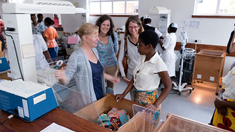
 Joe Langton [left], Maria Oden, and Rebecca Richards-Kortum talk to a new mother about the continuous positive airway pressure (CPAP) machine being used at Chatinkha Nursery in Blantyre, Malawi.
Richards-Kortum returned to Texas with a new mission: designing medical equipment for clinics in underserved communities that could withstand harsh climate conditions and be easily repaired. She also wanted to get students involved in the work.
To help her cause, she and colleague Z. Maria Oden, also a bioengineering professor, founded the Rice360 Institute for Global Health Technologies. Undergraduate and graduate students at the institute develop affordable medical technologies to help solve health challenges worldwide.
Richards-Kortum formed an institute team of researchers, physicians, and students to design a tool that could detect precancerous cells to help prevent oral and cervical cancer.
Precancerous cells, which have grown abnormally in size, shape, or appearance, have a high chance of becoming cancerous. Precancerous epithelial cells in the mouth and the cervix, in particular, are likely to develop into cancer. The most common sign epithelial cells are precancerous is that their nuclei are enlarged, according to the American Cancer Society.
When precancerous tissue forms, new blood vessels grow to supply it with blood. Because hemoglobin in the red blood cells absorbs visible light, Richards-Kortum's team developed a fiber-optic probe that can produce images of the underlying network of new vessels. The tool also can image epithelial cells and their nuclei.
The high-resolution micro-endoscope (HRME) provides answers about a person's intracellular structure without the need for a biopsy. The device, which is about the size of a DVD player, houses a 475-nanometer mirror, an optical sensor, and a 150-millimeter tube lens. Connected on one side is a flexible fiber bundle, just 1 mm in diameter, with a light source and a digital CCD camera inside. The light source is a blue LED with a peak wavelength of 455 nm. On the other side of the device is a cable that can be connected to a laptop, a tablet, or a smartphone.
To image a patient's tissue, a physician applies topical contrast gel to the area to be tested, then places the fiber bundle on the tissue. Some of the light from the fiber bounces back from the tissue, and those emissions are transmitted through the mirror and focused onto the optical sensor and the tube lens. Images of the epithelial cells are transferred to a laptop, tablet, or phone. The HRME can image the area at 80 frames per second. The device correctly identifies precancerous tissue 95 percent of the time, Richards-Kortum reports, and AI-based algorithms are being incorporated into the tool to further improve its performance.
By [using the tool] physicians can correlate the changes in nuclear structure and the changes in the vascular structure to see if there are a large number of precancerous cells," Richards-Kortum says. Health care workers are using the HRME to screen patients for cervical, oral, and esophageal cancer in clinics around the world, including in Botswana, Brazil, and El Salvador.
Improving neonatal care in sub-Saharan AfricaIn 2007 Richards-Kortum, Oden, and their team began developing technology to improve neonatal health care and reduce death rates in sub-Saharan Africa.
Their first invention was a continuous positive airway pressure (CPAP) machine for newborns with breathing problems. It consists of a shoe box that houses a 900-gram reusable water bottle, which is connected to a pump that sends air through the bottle and into the baby's airways. Their CPAP machine was commercialized in 2014 and is now being used in more than 35 countries.
But that tool helped with only one health issue newborns might face, she says. To develop medical devices to improve comprehensive care for newborns, she and Oden helped launch Newborn Essential Solutions and Technologies, known as NEST360, in 2017. The initiative brings together engineers, physicians, health care experts, and entrepreneurs from 12 organizations including the Malawi College of Medicine, the London School of Hygiene and Tropical Medicine, and the Ifakara Health Institute.
The initiative developed the NEST360 newborn tool kit. It includes 17 machines including a radiant warmer and incubator to help maintain an infant's body temperature; diagnostic tools for sepsis and infections; and a low-power syringe pump to dispense medicine, fluid, or formula. The group has trained 10,000 medical professionals on how to use the kits.
Today, 65 hospitals and clinics across Kenya, Malawi, Nigeria, and Tanzania are using the tool kits, which will soon be supplied to hospitals in Ethiopia, officials say.
NEST360 estimates that the kit is improving the lives of 500,000 newborns annually.
Being part of a team that is providing care to patients who have not been traditionally well served by our existing health system is a privilege," Richards-Kortum says.
A bridge between EE and health careRichards-Kortum joined IEEE while teaching at the University of Texas.
I really appreciate the way the organization has thought about the intersectionality between electrical engineering and health care technology," she says. IEEE has been an important voice in moving that field forward for faculty members and students, and doing that in a way that prioritizes equity."
Professional networking opportunities are also an important benefit, she says. Richards-Kortum recommends her students join IEEE not only for the networking avenues but also for the professional development and continuing education programs, as well as the ability to share and learn about advances in research.