Do-it-yourself breast ultrasound
Early detection is key to surviving breast cancer, but tumors that develop in between routine mammograms-known as interval cancers-tend to be especially aggressive. A wearable ultrasound device devised by MIT researchers could help detect such tumors when they are still in early stages.
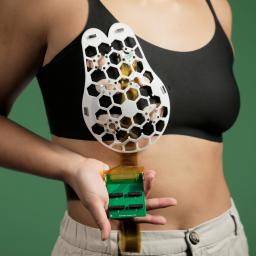
The device can be attached to a specialized bra to let an ultrasound tracker image the breast tissue from different angles. In their study, the researchers showed that they could obtain images comparable in resolution to those done at medical imaging centers.
We changed the form factor of the ultrasound technology so that it can be used in your home. It's portable and easy to use, and provides real-time, user-friendly monitoring of breast tissue," says Canan Dagdeviren, an associate professor in MIT's Media Lab and the senior author of the study. Dagdeviren drew up the first rough schematic of the device as an MIT postdoc at the bedside of her aunt Fatma Caliskanoglu, who (despite regular screenings) died of breast cancer six months after receiving a diagnosis at age 49.
My goal is to target the people who are most likely to develop interval cancer," says Dagdeviren, whose research group specializes in developing wearable electronic devices. With more frequent screening, our goal is to increase the survival rate to up to 98%."
To make her vision of a diagnostic bra a reality, Dagdeviren designed a scanner that's based on the same kind of technology used in medical imaging centers but can be much smaller thanks to the use of a novel piezoelectric material.
To make the device wearable, the researchers designed a flexible, 3D-printed patch, which has honeycomb-like openings. Using magnets, this patch can be attached to a bra with openings that allow the ultrasound scanner to contact the skin. The scanner fits inside a small tracker that can be moved to six different positions, and it can be rotated to take images from different angles. It does not require any special expertise to operate.
Working with the MIT Center for Clinical and Translational Research, the researchers tested their device on a 71-year-old woman with a history of breast cysts and succeeded in detecting cysts as small as 0.3 centimeters in diameter-the size of early-stage tumors. The patch can be used over and over, and the researchers envision that it could be used at home by people at high risk for breast cancer. It could also help diagnose cancer in people who don't have regular access to screening.
Today, the researchers have to connect the device to a traditional ultrasound machine to view the images. But they are working to develop a smartphone-size version of the system used to read ultrasound scans, so that patients wouldn't have to visit an imaging center.
Eventually, artificial intelligence might be used to analyze how the images change over time, which could be more accurate than relying on the assessment of a radiologist comparing images taken years apart. The researchers also plan to explore adapting the ultrasound technology to other parts of the body.