Google helped make an exquisitely detailed map of a tiny piece of the human brain
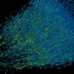
A team led by scientists from Harvard and Google has created a 3D, nanoscale-resolution map of a single cubic millimeter of the human brain. Although the map covers just a fraction of the organ-a whole brain is a million times larger-that piece contains roughly 57,000 cells, about 230 millimeters of blood vessels, and nearly 150 million synapses. It is currently the highest-resolution picture of the human brain ever created.
To make a map this finely detailed, the team had to cut the tissue sample into 5,000 slices and scan them with a high-speed electron microscope. Then they used a machine-learning model to help electronically stitch the slices back together and label the features. The raw data set alone took up 1.4 petabytes. It's probably the most computer-intensive work in all of neuroscience," says Michael Hawrylycz, a computational neuroscientist at the Allen Institute for Brain Science, who was not involved in the research. There is a Herculean amount of work involved."
Many other brain atlases exist, but most provide much lower-resolution data. At the nanoscale, researchers can trace the brain's wiring one neuron at a time to the synapses, the places where they connect. To really understand how the human brain works, how it processes information, how it stores memories, we will ultimately need a map that's at that resolution," says Viren Jain, a senior research scientist at Google and coauthor on the paper, published in Science on May 9. The data set itself and a preprint version of this paper were released in 2021.
Brain atlases come in many forms. Some reveal how the cells are organized. Others cover gene expression. This one focuses on connections between cells, a field called connectomics." The outermost layer of the brain contains roughly 16 billion neurons that link up with each other to form trillions of connections. A single neuron might receive information from hundreds or even thousands of other neurons and send information to a similar number. That makes tracing these connections an exceedingly complex task, even in just a small piece of the brain..
To create this map, the team faced a number of hurdles. The first problem was finding a sample of brain tissue. The brain deteriorates quickly after death, so cadaver tissue doesn't work. Instead, the team used a piece of tissue removed from a woman with epilepsy during brain surgery that was meant to help control her seizures.
Once the researchers had the sample, they had to carefully preserve it in resin so that it could be cut into slices, each about a thousandth the thickness of a human hair. Then they imaged the sections using a high-speed electron microscope designed specifically for this project.
Next came the computational challenge. You have all of these wires traversing everywhere in three dimensions, making all kinds of different connections," Jain says. The team at Google used a machine-learning model to stitch the slices back together, align each one with the next, color-code the wiring, and find the connections. This is harder than it might seem. If you make a single mistake, then all of the connections attached to that wire are now incorrect," Jain says.
The ability to get this deep a reconstruction of any human brain sample is an important advance," says Seth Ament, a neuroscientist at the University of Maryland. The map is the closest to the ground truth that we can get right now." But he also cautions that it's a single brain specimen taken from a single individual.
The map, which is freely available at a web platform called Neuroglancer, is meant to be a resource other researchers can use to make their own discoveries. Now anybody who's interested in studying the human cortex in this level of detail can go into the data themselves. They can proofread certain structures to make sure everything is correct, and then publish their own findings," Jain says. (The preprint has already been cited at least 136 times.)
The team has already identified some surprises. For example, some of the long tendrils that carry signals from one neuron to the next formed whorls," spots where they twirled around themselves. Axons typically form a single synapse to transmit information to the next cell. The team identified single axons that formed repeated connections-in some cases, 50 separate synapses. Why that might be isn't yet clear, but the strong bonds could help facilitate very quick or strong reactions to certain stimuli, Jain says. It's a very simple finding about the organization of the human cortex," he says. But we didn't know this before because we didn't have maps at this resolution."
The data set was full of surprises, says Jeff Lichtman, a neuroscientist at Harvard University who helped lead the research. There were just so many things in it that were incompatible with what you would read in a textbook." The researchers may not have explanations for what they're seeing, but they have plenty of new questions: That's the way science moves forward."
Correction: Due to a transcription error, a quote from Viren Jain referred to how the brain exports' memories. It has been updated to reflect that he was speaking of how the brain stores' memories.