New imaging tech gives us the finger (of a mummy)
Enlarge (credit: Romell et al. 2018)
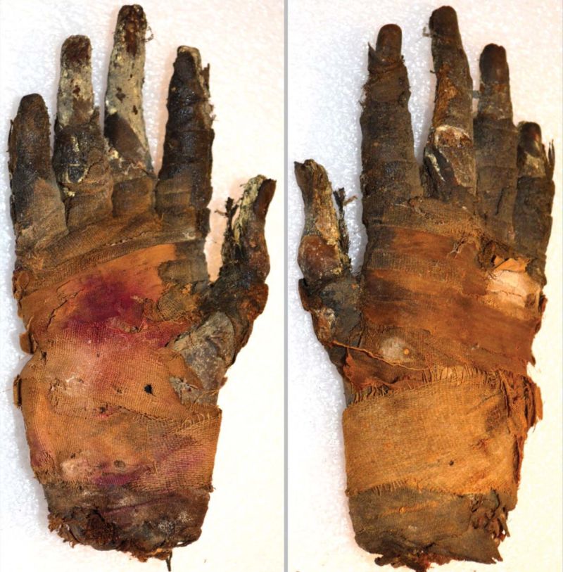
A variation on traditional CT scanning recently gave archaeologists a much closer look at a 2,400-year-old mummified hand from ancient Egypt. Medical imaging already uses the technology for higher-resolution views of patients' soft tissues, but this is the first time it has been tested on mummified human remains.
For soft tissue imaging, contrast is key"There has long been a struggle to retrieve information from ancient soft tissues, and neither conventional X-ray imaging and CT, nor new methods like MRI and terahertz imaging, have been able to give images with enough contrast or resolution for detailed analysis of these tissues," physicist Jenny Romell of the KTH Royal Institute of Technology in Sweden told Ars Technica.
CT scanning relies on X-rays but uses a computer to combine images from different angles into cross-sectional views of the body. Differences in the amounts of radiation absorbed by different materials produce the contrast visible in the resulting image. The method works especially well for hard, dense materials like bone, but most types of soft tissue don't produce enough contrast for really high-resolution imagery. Instead, paleopathologists who want to study ancient tissues have to take small samples from mummified remains and examine them under a microscope.
Read 8 remaining paragraphs | Comments