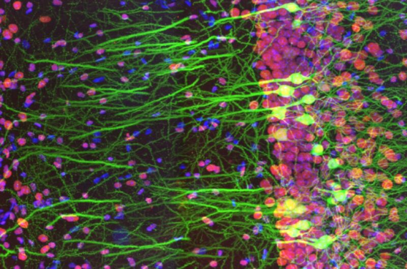
Extreme closeup of mouse-brain slice wins top Life Science Microscopy prize

Enlarge / Detail from the winning entry in the first Olympus Global Image of the Year Life Science Light Microscopy Award. It shows immunostaining of a mouse-brain slice with two fluorophores. (credit: Ainara Pintor/Olympus)
For several years now, we've regularly featured the winners of Nikon's annual Small World microscopy contest. Now, Olympus has entered the artful imaging arena with its first Global Image of the Year Award. Like the Small World contest, the intent is to highlight artful scientific imaging in hopes of inspiring the world to appreciate the inherent beauty of microscopy imaging. Olympus announced the winners (one global winner, plus three regional winners), along with several runners-up, last month. They do not disappoint.
As Ars' John Timmer noted in his 2018 Small World coverage: "Microscopy is a sibling of photography in many ways beyond the involvement of high-end lenses. While it might not matter for scientific purposes, a compelling microscope image depends on things like composition, lighting, exposure, and more. And these days, both fields rely heavily on post-processing." All those elements are abundant in the new crop of Olympus winners.
Spain's Ainara Pintor snagged the top honor from over 400 submissions with her gorgeous image of an immunostained mouse-brain slice, titled Neurogarden. The image focuses on the hippocampus area of a single slice, but there are more than 70 million neurons in the mouse brain as a whole, according to Pintor. Howard Vindin of Australia won the regional prize for Asia-Pacific by capturing an autofluorescence image of a mouse embryo. US entrant Tagide de Carvalho won the regional award for the Americas with his colorful image of a tardigrade. The regional winner for Europe, the Middle East, and Africa was the UK's Alan Prescott, for his image capturing the frozen section of a mouse's head.
Read 1 remaining paragraphs | Comments